CSF Biothermofluidics
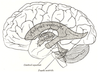
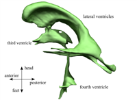
The cerebrospinal fluid (CSF) is a liquid of the body of many mammals. The fluid is colorless and it occupies the subarachnoid space and the ventricular system. The ventricles are four connected cavities in between parts of the brain. The subarachnoid space is the space between the brain and the skull. The brain therefore floats in the CSF and is protected very well against heavy accelerations. The CSF is produced in the choroid plexus, a sponge like structure situated in the ventricles. The CSF flows from the choroid plexus through the different ventricles and via an opening in the subarachnoid space where it is absorbed in the arachnoid villi and the fluid is fed back into the venous blood system. Upon this steady state flow, a pulsatile component is superimposed with the frequency of the heart beat. This pulsatile component is dominant compared to the steady state part. Many severe and potentially fatal diseases, such as e.g. hydrocephalus, normal pressure hydrocephalus or meningitis, are closely connected to the CSF. To improve diagnostics and treatment of diseases connected to the CSF, an improved knowledge of the functional mechanisms is desired. One major challenge is the integrated nature of all organs in the human skull which makes invasive measurements very risky and sometimes even unreliable.
Therefore non-invasive ways or phantoms are crucial to gain more knowledge.
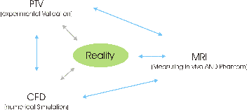
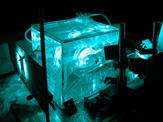
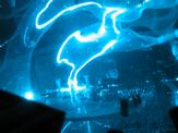
Within the framework of this thesis, different aspects of the dynamics of the CSF were investigated. Using a flow phantom with very accurately reproduced geometry, a detailed insight into the instationary flow field of the third ventricle has been provided using particle tracking velocimetry (PTV) and magnetic resonance imaging (MRI) techniques.
This project is part of a Polyproject, a collaboration among many institutes and universities (LTNT, IBT, BIWI (ETH), Neuroradiology (USZ), University of Oxford)
Within the interdisciplinary collaboration the various participants contribute their own expertise. For details about the work of the other members of the team, please follow the links in the summary.
In the vast variety of modeling approaches, there has been a long debate about how the motion of the CSF is driven. Generally accepted has been the assumption that the motion of the CSF is driven indirectly by the pulsatile pressure of the cranial arteries. Modeling this energy transfer is challenging. First, there is no obvious causal chain or ordered sequence of events. Secondly, access to the energy transfer via measurements is difficult. The motions of the brain tissue, ventricle walls, choroid plexus, etc. are very small and difficult to measure, even with state-of-the-art magnetic resonance imaging (MRI) methods. There are three main hypotheses explaining the driving force of the (pulsatile) CSF flow. One of the early attempts at explaining the propulsion is a "thalamic pump" is proposed as the origin of the CSF motion. Based on observations of CSF motion, its author concludes that the driving force consists of the two thalami squeezing together the third ventricle and thus pumping the cerebrospinal fluid.
Other publications claim that the CSF flow is pumped by the oscillating motion of the brain or directly by the displacement of the ventricular walls. This very motion of either tissue must be driven by intracranial arteries, both of which suggest that the driving force originates in the intracranial arteries. Brain motion is usually measured via MRI techniques.
The third hypothesis suggests that the CSF is driven by a pulsating choroid plexus. This hypothesis is supported by the fact that the choroid plexus is highly vascularized.
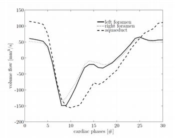
With measurements of the volume flow in the foramina of Monro and the aqueduct of Sylvius in several volunteers we have shown that the magnitude of the volume flow in the foramina of Monro and the aqueduct of Sylvius are of the same order of magnitude. Peak velocities in the foramina of Monro are smaller than in the aqueduct of Sylvius. The phase difference between the flows of the aqueduct of Sylvius and the foramina of Monro shows a significant inter-subject variability. The presented mathematical model with a propulsion by the choroid plexus is capable of explaining the phase shifts and their variances. Therefore, the measurement results confirm the validity of the model of the propulsion of the CSF via the choroid plexus.
"In-vitro measurement of ventricular cerebrospinal fluid flow using particle tracking velocimetry and magnetic resonance imaging", M. Schibli, M. Wiesendanger, L. Guzzella, K. Hoyer, M. Soellinger, V. Kurtcuoglu, P. Boesiger; 2008 First International Symposium on Applied Sciences on Biomedical and Communication Technologies (ISABEL), IEEE
"Investigations of Ventricular Cerebrospinal Fluid Flow Phase Diferences between the Foramina of Monro and the Aqueduct of Sylvius", M. Schibli, M. Wyss, P. Boesiger, L. Guzzella. Biomedizinische Technik